Glaucoma treatment in Ukraine
Glaucoma is a rapidly progressing pathology, leading to the onset of complete blindness. According to WHO statistics, more than 14% of people who have lost vision had this serious disease.
Important! At the initial stage of the development of the disease, the intraocular pressure rises in patients, causing damaging of the optic nerve, which cannot be restored in the future. Sensory functions deteriorate, visual acuity is significantly reduced, and a loss of the field of view is observed.
Every year in Ukraine, this dangerous pathology is diagnosed in 20 thousand people.

Want to know how much the treatment costs?
Answer a few questions and get preliminary information about the cost of diagnosis and treatment!
Causes of glaucoma

The main factor causing the development of disorders is intraocular hypertension, which manifests itself in an increase in intraocular pressure. Common reasons of intraocular hypertension are:
- Fetal developmental anomalies. Congenital disorder occurs in about 1 case in 10,000;
- Genetics. In some cases, the predisposition to glaucoma is inherited;
- Visual impairment. Hyperopia can be detected in the closed-angle form of glaucoma, when the intraocular fluid drainage channel is closed by a significantly dilated pupil (the effect of drugs, darkness). This form progresses rapidly, observed in 10% of cases. The cause of myopia may be the open-angle form of pathology, when the drainage of moisture is disturbed in the open outflow channel. The progression is slow, detected in 90% of cases;
- Thin cornea. This physiological feature is observed in a large number of patients;
- Mechanical damage. The process of circulation of intraocular humid is disrupted due to head injuries (for example, due to contusion) or eyes (injuries, burns);
- Eye diseases. Inflammatory ophthalmic diseases (keratitis), as well as dystrophic changes (vitreous hemorrhage, iris atrophy);
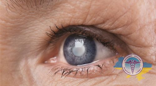
- Cataract. Phacomorphic pathology, as a result of which moisture removal is disturbed due to the swelling lens;
- Postoperative complications. The painful condition develops after improperly performed interventions or as a result of complications arising after ophthalmic surgery;
- Diabetes. The consequence may be the development of severe diabetic retinopathy, causing damage in the small vessels of the fiber;
- Hypertension. Pathology can develop due to high blood pressure.
What are the symptomatic manifestations of glaucoma?
The disease is practically not amenable to therapy; after destruction of optic nerve it cannot be restored. But it is possible to save visual functions or significantly slow down the process with early diagnosis. The disease has no distinct stages. At an early stage, dark spots appear when looking, vision deteriorates, which is not immediately noticed by patients. The surgery at this stage helps to normalize the pressure.
In the process of further development, peripheral vision decreases - a person recognizes only what is located directly in front of him, the lateral vision deteriorates. At this stage patient usually turns for help.
The last stage is terminal, when the treatment does not give results and the person is completely blind.
The following symptoms are distinguished:
- Pain that occurs locally or throughout the head;
- The appearance of sharp pain in the eyes, floating dark spots, blurred eyes;
- In the evening and at night, redness of the eyes is observed; in low light, objects are less distinguished;
- When looking at light sources (fire, lamp), halos and a rainbow appear;
- Narrowing of the field of view, the occurrence of peripheral vision problems;
- Frequent occurrence of nausea and vomiting.
Attention! In many cases, the painful condition proceeds without pronounced signs, the deterioration of the condition is not noticed immediately. Early detection is facilitated by regular screening examinations, since the symptoms that are obvious to the patient himself are clearly manifested when the damage to the nerve fibers has already reached more than 40%.
What are the types of glaucoma?
Depending on what caused the increase in intraocular pressure, the following types of glaucoma are distinguished:
- Open-angle glaucoma. Development occurs gradually, due to which there are practically no obvious symptoms and warning signs in the early stages. A person notices a deterioration in vision, a narrowing of vision, "blind spots" appear and he or she is poorly oriented in space, when the process is already making significant progress and more than 40% of nerve fibers have been lost;
- Closed angle. Sudden worsening of the condition due to blockage of the drainage system. In the case of complete closure of the corner, an acute attack occurs, which can imitate other conditions: headache and toothache, intestinal upset, flu, nausea, and can also manifest itself in the form of general weakness. Complete blindness can develop within a few days or even hours, so in case of an attack, you must immediately consult an ophthalmologist;
- Normal pressure glaucoma. The onset of the disease is possible with statistically normal IOP values. In these situations, the pressure has a negative effect and damages the nerve fibers;
- Secondary neovascular. The source of the onset of the painful condition is the newly formed vessels that block the drainage system. This may be a consequence of PCV thrombosis, diabetes mellitus and other disorders in the body;
- Ophthalmic hypertension. It manifests itself in the form of increased IOP, while additional examination does not reveal the presence of nerve damage, and there are also no typical changes in the visual field.
How is glaucoma diagnosed in Ukrainian clinics?
Eye disease can take place in 4 stages, manifesting itself at the initial stage and ending with the terminal one. Most people begin to notice symptoms only at an advanced stage or already at a far advanced stage. Some signs (nausea, pain and dizziness) are often not equated with visual impairment.
Important! Timely diagnosis of glaucoma in Ukraine makes it possible to identify typical signs even before clinical symptoms appear clearly and clearly.
The main methods of ophthalmic diagnostics are:
- Tonometry. IOP indicators are measured using devices (contact, non-contact);
- Gonioscopy. The doctor examines the anterior chamber of the sensory organ, as well as the angle that captures the iris and cornea;
- Quantitative static perimetry. Using static objects of different sizes and having variable brightness, the doctor examines the topography of the field. Thus, different parts of the retina are examined for light sensitivity;
- Optical coherence tomography. The diagnostic method is applied to the retina and optic nerve. The anterior segment of the sensory organ is examined with an accuracy of a thousandth of a millimeter;
- Optical topography. Study of the state of the drainage system in the anterior segment, as well as measurement of the thickness of the cornea;
- Tonography, sphygmography. A survey that allows you to study the blood circulation process, as well as the permeability of moisture (its removal and inflow);
- Ultrasound biometrics. The technique allows you to study anatomical parameters, including the size of the eyeball, lens, etc.;
- Photo registration. With the help of special equipment, a snapshot of the nerve is taken, stating its real state.
How is therapy carried out in glaucoma clinics?
The technique is aimed primarily at reducing IOP. Depending on the type of disease and its severity, the following methods are used in glaucoma treatment clinics:
- Drug therapy. After examining and determining the nature of the violations, the doctor prescribes eye drops, the pharmacological action of which normalizes IOP;
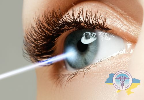
- Laser surgery. Intervention is prescribed in situations where the use of drops is ineffective or as an adjunct to drug therapy. After exposure to a high-precision beam, the outflow of moisture improves;
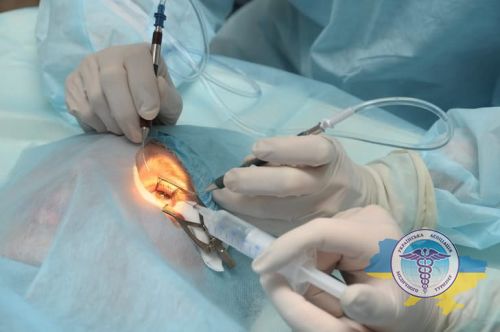
- Microsurgery. In the course of this surgical technology, a drainage channel is created, through which fluid is removed, which helps to reduce IOP.
Before prescribing a specific method, a thorough diagnostic examination is carried out, which makes it possible to identify the disease, as well as to determine its form, stage and concomitant pathologies. Based on the results of the research, an individual scheme for eliminating violations is assigned.
In the early stages, conservative methods are used, but drug therapy in the form of drops does not always achieve the desired effect.
At later stages, laser exposure or surgical techniques are required, which can be performed on an outpatient basis, therefore, hospital stay is not necessary.
Laser glaucoma treatment

One of the safest and most widespread methods in modern medicine is laser treatment of glaucoma, which shows effective results. It has a number of advantages: the procedure is accompanied by low trauma, patients in most cases do not have serious postoperative complications, manipulations are carried out on an outpatient basis, so no hospital stay is required.
Laser exposure is subdivided into the following types:
Trabeculoplasty
This technique is the "gold standard" among the various available methods of laser exposure. Under the action of the beam on the drainage zone (the area in which moisture is removed), certain segments of it expand.
As a result, outflow improves, IOP normalizes, and it stabilizes. To carry out the manipulations, no preoperative preparation is required, anesthesia is local (drip).
Sclerotomy
Good alternative for trabeculectomy. The procedure involves puncturing the sclera (outer shell) with a directional carbon dioxide beam. The technology minimizes the number of side effects and contributes to the normalization and stabilization of IOP.
Iridectomy
The technology is widely used in the closed-angle form, in which the size of the angle between the cornea and the iris is narrowed, which blocks the drainage of moisture, which, in turn, increases the pressure.
During iridectomy, a targeted high-precision beam produces an optimal opening in the iris, improving the outflow patency, thereby reducing pressure in the optic organ.
Surgical treatment of glaucoma in Ukraine

The intervention is prescribed in case of unstable IOP, when therapy and laser exposure do not achieve the desired result, or when these technologies are no longer applicable at later stages. For the treatment of glaucoma in Ukraine, the following methods are common:
Sclerectomy
The specified surgical intervention is of the non-penetrating type, i.e. with non-penetrating deep sclerectomy, opening of the eyeball is not required. In the process of manipulations, through holes are not used - a special valve is created in the sclera, allowing moisture to circulate unhindered, normalizing its outflow.
The balance is restored, IOP normalizes. After procedure the duration of the rehabilitation period is several days.
Viscocanalostomy
This technology differs from the NGSE in that the outflow is restored as a result of the introduction of a special viscous substance - Gealon (Artiviska) into the drainage channels, the lumen of which expands. Gealon also helps to restore moisture permeability and regulates its balance without directly opening the apple. The rehabilitation period lasts for 1-2 days.
Implantation of the Ex-PRESS device
One of the latest technologies that is popular in the United States and the European Union. After the procedure, the physiological pathways for moisture removal are restored. During the manipulations, a specialized Ex-PRESS device manufactured by the American company Alcon is implanted.
The use of an implantable device is a safe method of lowering IOP with minimal postoperative complications. Available technology quickly and effectively relieves of a dangerous ailment, regardless of neglect and severity.
Installing the Ahmed Valve
In the modern technique, the Ahmed valve is implanted - a special device whose purpose is to reduce IOP and establish control over it. As a result of implantation, moisture moves freely outward from the anterior chamber, which provides a decrease in pressure.
The valve is a drainage system, the main element of which is a unique membrane that opens the device in case of excess pressure in the front chamber. On the back surface of the valve, there are special tubes that ensure the connection of the subtennon space with the anterior chamber of the sensory organ, allowing unhindered humid removal.
What determines the price of glaucoma treatment in Ukraine?
The cost of glaucoma treatment in Ukrainian clinics is influenced by the applied methodology: with the conservative method, the prices for drugs and the duration of the prescribed course are taken into account, with the operative method, the type of intervention selected according to the results of the examination, taking into account contraindications.
Stable prices for laser glaucoma treatment depend on the chosen technique and the equipment used. Surgical intervention is technologically more difficult to perform, therefore it is more expensive than laser therapy.
The structures that are affected during the procedure are also taken into account, for example, the iris or the trabecula. Ciliary body manipulations are less common, which increases the cost of the procedure.
Depending on the clinic, the average cost of laser treatment ranges from $ 500 to $ 1000. Surgical intervention or implantation of the Ex-PRESS device - from $ 1500.


